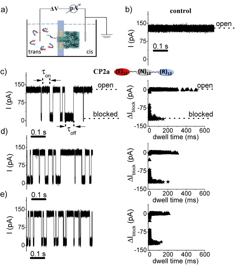
Figure 1.
Detecting single polypeptides with a nanopore. a) Schematic illustration of the apparatus (note that α-HL, peptides and chamber are not drawn to scale). The pore is formed by and the voltage is applied across the pore via a matched pair of Ag-AgCl electrodes. b) Ionic current time series of the open pore. Addition of the CP2a peptide at c) 5 μM, d) 10 μM, or e) 20 μM to the trans side causes randomly occurring transient blockades in the current. The applied potential was +70 mV (a positive voltage drives cations from the trans to the cis compartment) and the solution contained an aqueous electrolyte solution of 2 M KCl, 10 mM HEPES, at pH = 7.3.

