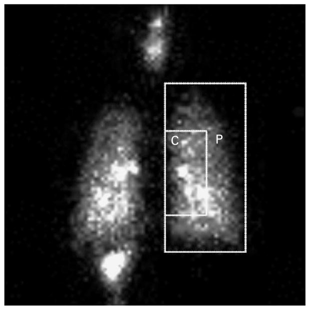
Figure 2.
Posterior gamma camera image of the whole lung showing regions used for determination of the central to peripheral (C/P) ratio and to evaluate the regional deposition of inhaled particles. The larger rectangle outlines the whole right lung as defined by a xenon equilibrium scan, while the smaller rectangle defines the central region (C), comprised of a higher proportion of large airways relative to the lung periphery (P). The very bright areas within the lung represent radioactivity concentrated in central airways.

