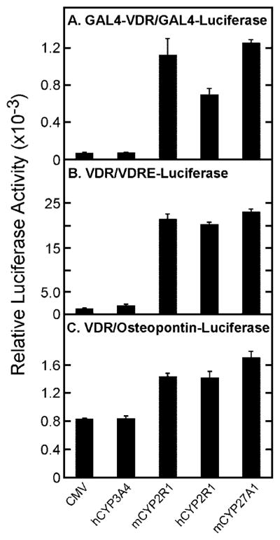
Fig. 3. Activation of vitamin D3 by CYP2R1.

The indicated vitamin D receptor-reporter systems were introduced into HEK 293 cells grown in 60-mm dishes together with the expression plasmids designated at the bottom of the figure. Relative luciferase enzyme activity was determined 16–20 h later as indicated under “Experimental Procedures,” and the means ± S.E. for experiments carried out in triplicate were plotted as histograms. The amounts of vitamin D3 added to the medium were 0.5 μM (A and B) and 1 μM (C).
