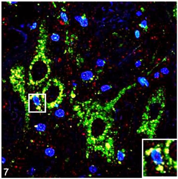
Figure 7.
Parasympathetic nucleus of the vagus; cat, secondary passage FelCWD, case No. 1. Finely granular to clumped misfolded prion protein (PrPD) deposits (red) are seen dispersed through the tissue, and dual immunofluorescence with the anti–lysosome antibody Lamp1 (green) reveals extensive accumulation of PrPD with lysosomes (yellow). The yellow color represents the overlapping of red and green immunofluorescent staining, and nuclei are stained with 4′,6-diamidino-2-phenylindole, dihydrochloride (DAPI; blue). Inset: higher magnification further illustrates the association between PrPD and Lamp1-positive lysosomes.

