
Figure 1:
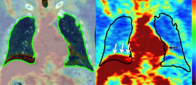
Left: Coronal section from pre-RT FDG PET/CT image, with the lung ROI segmented from CT (green on left, black on right). Right: Manual editing is required to remove the liver (white arrows) and heart (black arrow) from the lung ROI due to motion-induced misalignment of PET and CT.