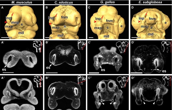
Fig 1.
Primary palate initiation in amniotes documented with OPT scanning. Frontal views of isosurfaces created in amira (A-D) and virtual OPT slices (A′-D″) of embryos are shown for E11.5 mouse (Mus musculus), ∼ 10-day crocodile (Crocodilus niloticus), stage 28 chicken (Gallus gallus) and stage 4 turtle (Emydura subglobosa). All embryos have open external nares (red arrows with tails in A-D). The nasolacrimal groove which separates the lateral nasal prominence from the maxillary prominence (red arrowheads in A-D) is used as a reference landmark to compare the site of initial fusion between facial prominences. In the mouse and crocodile embryos (A,B), fusion occurs jointly between the lateral nasal, medial nasal and maxillary prominences and the fusion point is approximately at the level of the nasolacrimal groove. In the chicken embryo (C) fusion begins between the frontonasal mass and the maxillary prominence inferior to the nasolacrimal groove. In the turtle (D), fusion initiates between the frontonasal mass and the lateral nasal prominence and the fusion zone is superior to the nasolacrimal groove. Optical sections in the anterior frontal plane show that the tips of the prominences are fused in all specimens, as demonstrated by the presence of a bilayered epithelial seam (A′,B′,C′,D′; white arrow), whereas posterior sections show closed choanae in mouse and crocodile and open choanae in the chicken and turtle (A″,B″,C″,D″; white arrowheads). Insets show the plane of section with a red line (A′,A″,B′,B″,C′,C′,D′,D″). Key: e, eye; es, epithelial seam; fnm, frontonasal mass; lnp, lateral nasal prominence; md, mandibular prominence; mnp, medial nasal prominence; mxp, maxillary prominence; s, stomodeum. Scale bars: 500 μm.

