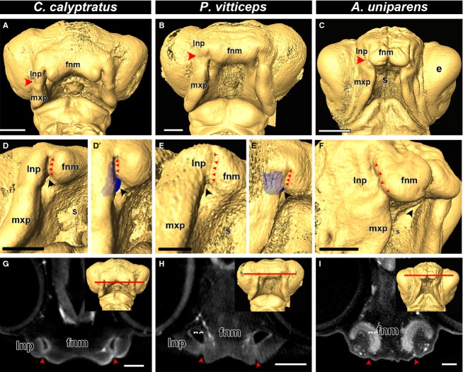
Fig 2.
Comparison of fusion in lizard embryos. Isosurfaces of stage 34 chameleon (Chamaeleo calyptratus), stage 31 bearded dragon (Pogona vitticeps) and stage 12 whiptail lizard (Aspidoscelis uniparens). Based on the position of the nasolacrimal groove (A-C; red arrowheads), primary palate development is initiated by the fusion of the frontonasal mass with the lateral nasal prominence. A closer view of the fusion zone reveals complete fusion of the lateral nasal prominence and the frontonasal mass, completely closing off the external nares (D,D′,E,E′,F; red arrowheads). An endocast of the nasal cavity (blue) reveals that it lies behind the fusion zone (D′,E′). Virtual coronal sections in the plane of the fusion zone (G,H,I; red line in the inset) reveals that the prominences are indeed fused to each other (G,H,I; red arrowheads) and there is an open cavity (the nasal cavity) directly behind the fused prominences. Key: e, eye; fnm, frontonasal mass; lnp, lateral nasal prominence; mxp, maxillary prominence; nc, nasal cavity; s, stomodeum. Scale bars: 500 μm.

