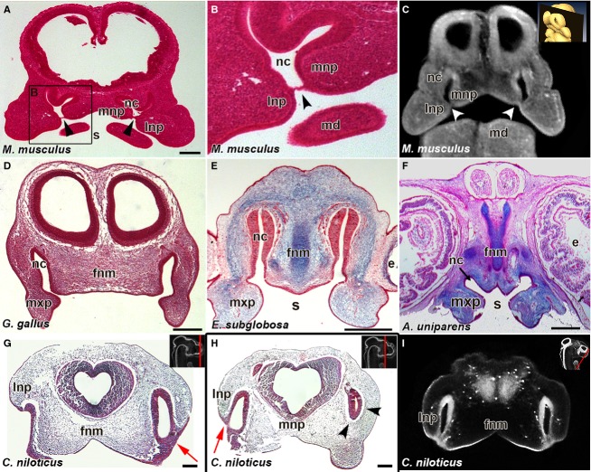
Fig 3.
Bucconasal membrane is only present in the mouse. Histological sections in the frontal plane from E11.5 mouse (Mus musculus) (A,B) and virtual section of E11.5 mouse (C), show the bucconasal membrane closing off the choanae from the stomodeum (black arrowheads in A, B; white arrowheads in C). In stage 28 chicken (Gallus gallus) (D), stage 4 turtle (Emydura subglobosa) (E) and stage 12 whiptail lizard (Aspidoscelis uniparens) (F) there is a continuous connection between the nasal cavity and the stomodeum via open choanae. Crocodilus niloticus embryo shows fused lateral and medial nasal prominences externally with an epithelial plug in between (red arrows, G,H). Posterior section reveals the region past the nasal fin on the left side of the head where mesenchyme surrounds the nasal cavity (H, arrowheads). Virtual Frontal section from OPT scan of 10-day crocodile embryo revealing fusion regions between the lateral nasal prominence and the frontonasal mass (I). Key: e, eye; fnm, frontonasal mass; lnp, lateral nasal prominence; mnp, medial nasal prominence; nc, nasal cavity; s, stomodeum. Scale bars: 250 μm (A); 200 μm (D-H).

