
Fig 5.
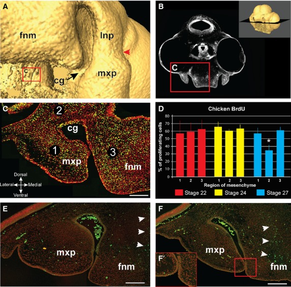
BrdU labeling in chicken embryos at the time of choana formation. A stage 27 embryo shows the opening between nasal and oral cavities (A). The coronal plane of section for BrdU cell counts passes through the choanal groove (B). (C) A representative section that passes through the maxillary prominence and frontonasal mass. Mesenchymal nuclei are labeled in red, BrdU-labeled cells are yellow. Percentage proliferating cells was calculated in three regions of mesenchyme (C); region 1 – maxillary prominence; region 2 – the base of the choanal groove; region 3 – the lateral edge of the frontonasal mass. At stage 22 and 24, where the choanal groove has not formed yet, there is no significant difference in proliferation among the three regions. However, at stage 27 there is a significant decrease in proliferation in region 2 (asterisk, P < 0.005) compared with the other regions, where proliferation rates remain approximately the same as in stages 22 and 24 (∼ 60%) (D). TUNEL analysis on stage 27 (E) and stage 28 (F) reveals minimal apoptosis in both the mesenchyme of the choanal groove (red arrowheads) as well as the fusion zone between the prominences (F′). The only region with significant apoptosis was the middle of the frontonasal prominence, where the nasal cartilage will form (white arrowheads). Key: cg, choanal groove; fnm, frontonasal mass; lnp, lateral nasal prominence; mxp, maxillary prominence, Scale bars: 100 μm (C,E,F).