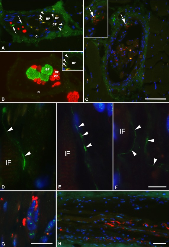
Fig 4.

Immunofluorescence micrographs of human muscle spindles of the deep neck muscles. (A and B) Two consecutive sections of a muscle spindle in the far A region. The NF-positive (red) spindle nerve contains TH-immunoreactive axons (green, arrow). Around both the MYH7b-positive (green) bag fibers (BF) and A4.74-positive (red) chain fiber (CF), very fine but specific labeling with TH (arrowheads) is present and shown at higher magnification in the inset in B. (C) Cross-section of a muscle spindle in the A region showing TH-positive axon (green, arrow) within a spindle nerve alongside with axons labeled with NF (red). In the inset in C, the spindle nerve is shown at higher magnification. (D–F) Longitudinally running axons, with varicose morphology and strongly labeled with the antibody against TH (green, arrowheads) are shown running in parallel with intrafusal fibers (IF) along different parts of the A region of one single muscle spindle from the deep neck muscles. (G and H) Annulospiral endings, strongly labeled with the antibody against NF (red), in the same muscle spindle as above, are shown for comparison. Nuclei are labeled blue with DAPI. C, capsule. Scale bar: 50 μm (A–C and H); 10 μm (D–F); 25 μm (G).
