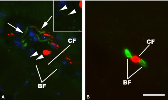
Fig 5.

Immunofluorescence micrographs of a human muscle spindle of the deep neck muscles. Two consecutive cross-sections in the C region, double-stained for demonstration of TH (green) and NF (red) (A) and MyHC slow tonic (green) and MyHC fast (red) (B). The C region fibers – two MyHC slow tonic positive bag fibers (BF) and one MyHC fast positive chain fiber (CF) – are closely associated to a TH- and NF-positive nerve (arrows). Very fine but specific labeling with TH (arrowheads) is present on the surface of a BF and shown at higher magnification in the inset. Scale bar: 25 μm.
