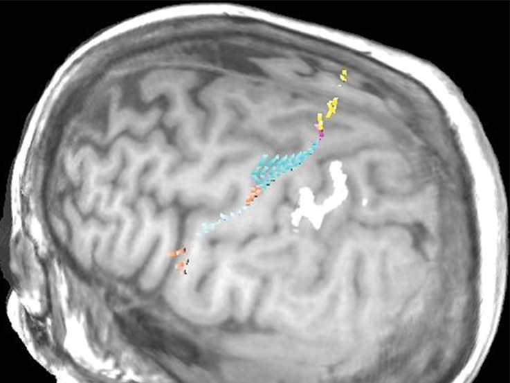
Figure 10d.

Normal variations in the precentral gyrus (purple shading), demonstrated by confocal volume rendering with an embedded deformable anatomic template of the corticospinal fibers and superimposed functional MR images of a subject performing a right-hand motor task. The fibers appear as dashed lines with color coding corresponding to that in Figure 6. White dot = hand knob, peach shading = potential cortical bridge. (a–c) Images show a single, roughly horizontal, T-shaped gyrus interrupting the precentral sulcus. (d–f) Images show a single interruption in the length of the precentral sulcus. (g–i) Images show two interruptions of the precentral sulcus.
