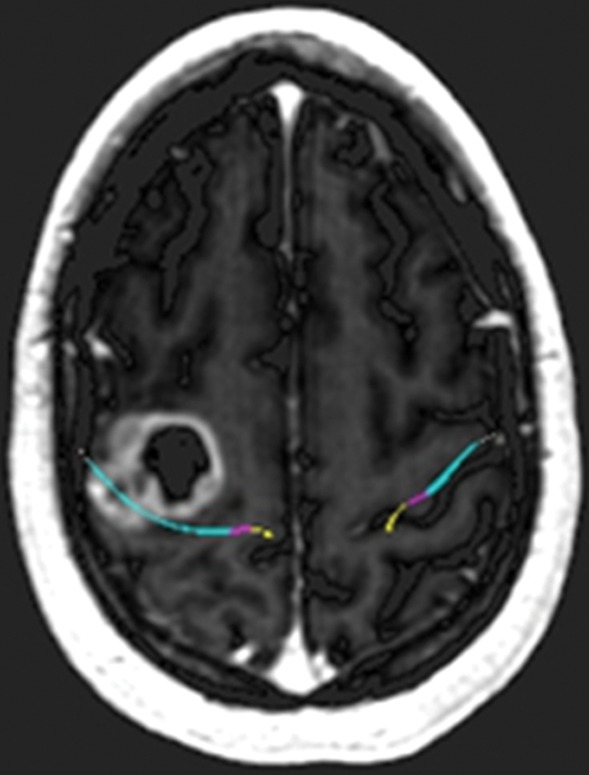
Figure 11a.

Glioblastoma under the hand knob. A 52-year-old woman noticed left third- and fourth-digit weakness that progressed to complete loss of left-hand function over 4–6 weeks. She was not a good candidate for functional MR imaging because of poor motor function. Diffusion tensor imaging was performed, but the tracts were not well seen. Contrast-enhanced T1-weighted MR images with embedded deformable anatomic template (see Fig 6 for color coding key) are helpful to show the relationship to the motor tract. The atlas-based motor tract was deformed around the glioblastoma to show the possible deviation in the tracts, assuming the tracts were intact. Resection of the rim-enhancing lesion under the hand knob revealed glioblastoma. After surgery, the patient had dense left hemiplegia, which improved slightly over 8 months of physical therapy. (a) Axial contrast-enhanced T1-weighted MR image shows how the tracts may be displaced posterior to the tumor. (b) Sagittal contrast-enhanced T1-weighted MR image shows the manual deformation of the upper extremity tracts and the normal course of the lower face and tongue motor fibers.
