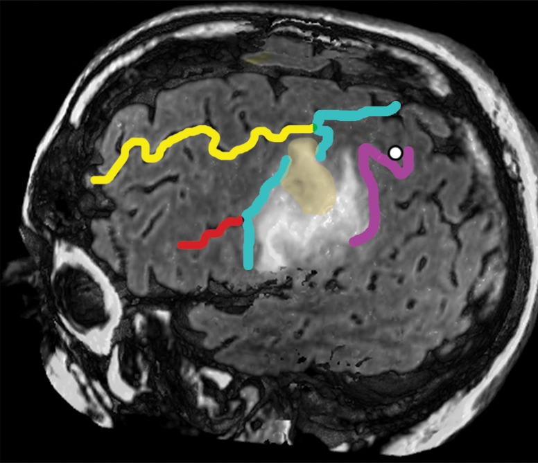
Figure 13b.

Confocal reconstructions of FLAIR MR images obtained in a 49-year-old man who presented after a grand mal seizure show an infiltrative lesion. Biopsy performed because of motor cortex involvement revealed anaplastic oligodendroglioma. Resection was terminated when intraoperative stimulation produced a contralateral facial seizure. (a) View of the opercular region shows involvement of BA 4 and BA 6 on the expanded precentral gyrus. Dark green = sylvian fissure outlining pars opercularis (BA 44), light green = sylvian fissure outlining pars triangularis (BA 45). (b) View of the posterior middle frontal gyrus shows infiltration of oligodendroglioma over the cortical bridge (peach shading) caused by interruption in the precentral sulcus. White dot = hand knob.
