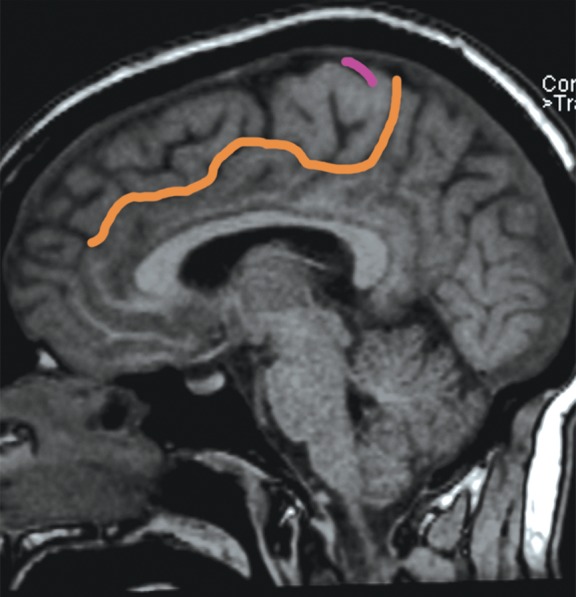
Figure 8a.

The bracket sign. Once the marginal ramus (orange) is located, the central sulcus (purple) is identifiable as the first sulcus immediately anterior to the marginal ramus. (a) Sagittal unenhanced T1-weighted MR image. (b) Axial unenhanced T1-weighted MR image.
