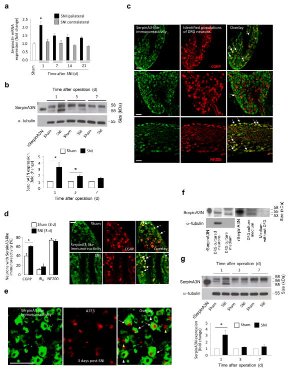
Figure 2.
Serpina3n is upregulated in mouse lumbar DRGs following spared nerve injury, a model of neuropathic pain. (a) Real time quantitative PCR (qPCR) analysis of Serpina3n expression in L3–L5 DRGs post-SNI, Cyclophilin A serving as reference gene (n = 4 mice per time point; *P < 0.05 compared to sham DRG; 1-way ANOVA, Tukey’s post-hoc test). (b) Western blot analysis and quantification of SerpinA3N expression (55 kDa band) in L3–L4 DRGs post-SNI normalized to α-tubulin expression (n = 3–5 mice/ time point,1 way ANOVA, Tukey’s post-hoc test). (c) SerpinA3-like immunoreactivity in peptidergic (CGRP) and non-peptidergic (IB4-binding) nociceptors and large diameter neurons (NF200) in L3–L4 DRG sections from naïve mice (arrows indicate areas of colocalization). (d) Quantitative analysis of SerpinA3-like immunoreactivity in L3–L4 DRGs 3 days post-SNI as compared to sham (n = at least 3 DRG sections/mouse, 3 mice/treatment group; *P < 0.05 compared to sham; two-tailed unpaired T-test) and examples of upregulation in the CGRP-expressing population. (e) SerpinA3 and ATF3 staining in L3–L4 DRGs at day 1 post-SNI showing SerpinA3+/ATF3+ (white arrows), Serpina3+/ATF3− (white arrowheads) and SerpinA3−/ATF3+ (yellow arrowheads) neurons. (f) Western blot analysis of SerpinA3N expression in lysates and medium from cultured DRG neurons. (g) Western blot analysis and quantification of SerpinA3N expression in lumbar spinal cord post-SNI (n = 3–5 mice/time point, *P < 0.05 compared to sham, 1-way ANOVA, Tukey’s post-hoc test). Scale bars represent 100 μm in (c), (d) and (e). Error bars: standard error of mean.

