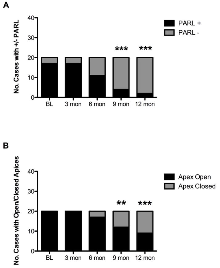
Figure 1.
Radiographic findings regarding the proportion of subjects with periapical radiolucencies and open apices at various time points of the study. A) The proportion of subjects with a periapical radiolucency decreased throughout the study period and became significantly different from baseline after 9 months (McNemar’s chi2 test, 6 months: p= 0.06; 9 and 12 month: p<0.0001). B) The proportion of subjects with an open apex began to decrease after 6 months, and became significantly different from baseline at 9 months (McNemar’s chi2 test, 9 months p<0.05; 12 months p<0.001).

