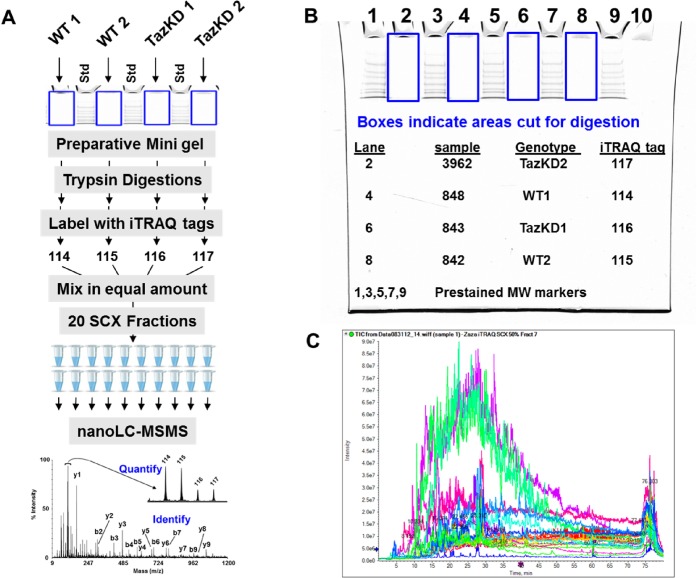
Fig 1. iTRAQ workflow for differential protein profiling in mitochondria from 3-month-old wild type and TazKD mice.
(A) Sample preparation workflow from a preparative SDS-PAGE gel, trypsin digestion, iTRAQ labeling, fractionation by strong cation exchange, followed by nanoLC-MS/MS to produce both protein identification by peptide fragmentation data and relative quantitation from the iTRAQ reporter fragment ions. (B) The preparative SDS-PAGE gel with pre-stained molecular weight markers showing the gel regions collected for trypsin digestion. (C) Overlay of the nano-LC-MS total ion current (TIC) from each of the 20 SCX fractions.

