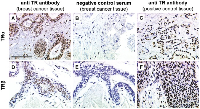
Fig 1. TRα and TRβ immunostaining.

Positive TRα (A) and TRβ (D) staining was detected in breast cancer tissue. Negative (TRα (B) and TRβ (E)) and positive controls (TRα (C) and TRβ (F)) were performed to validate staining specificity. Thyroid gland and vaginal epithelium served as tissue positive controls for TRα (C) and TRβ (F), respectively. Scale bar represents 100 μm and applies to A-F. Representative photomicrographs are shown.
