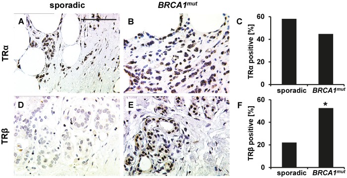
Fig 2. TRα and TRβ staining in breast cancer tissue.

Representative photomicrographs of TRα and TRβ immunohistochemistry staining in breast cancer tissue are shown. TRα was found to be abundantly expressed though there was no significant difference regarding the number of TRα positive cases when sporadic (A) vs. BRCA1 (B) mutated cancers were compared (C). Representative images of a TRβ staining scored as negative (D) and a TRβ staining scored as positive (E) in spontaneous (D) and BRCA1 mutated (E) cancer tissue are presented, respectively. TRβ was found to be expressed more frequently in BRCA1 mutated (E) cases as compared to spontaneous breast cancers (D). The fraction of TRβ positive cases in each group is illustrated in F. Significant changes are indicated by stars (*) and scale bar (applies to A, B, D and E) represents 100 μm.
