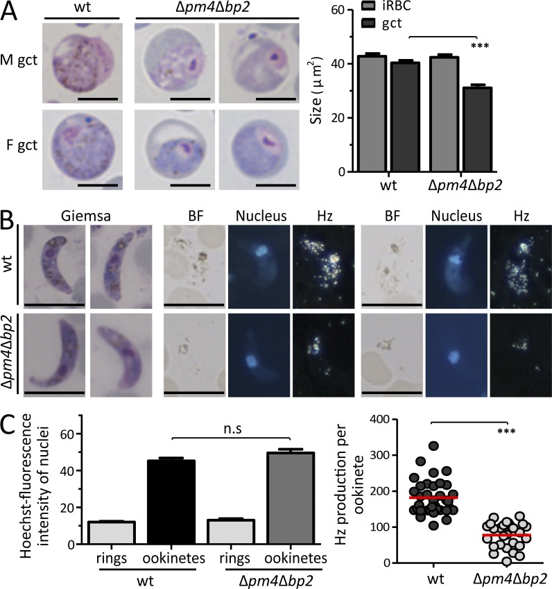
Figure 4.
Gametocytes of Δpm4Δbp2 are fertile despite their smaller size and reduced Hz production. (A) Representative Giemsa-stained images of WT and Δpm4Δbp2 gametocytes (left; bars, 5 µm) and the size quantification on these images (right; n > 30; ***, P < 0.0001; Student’s t test). (B) Giemsa-stained images and polarized light microscopy images of ookinetes. BF, bright-field; Nuclei (blue) stained with Hoechst. Bars, 5 µm. (C) Hoechst fluorescence intensity (DNA content) of WT and Δpm4Δbp2 ookinetes, relative to haploid ring-form stages, was quantified (left; n > 25; n.s, not significant; Student’s t test) indicating that both WT and Δpm4Δbp2 ookinetes are tetraploid. Relative light intensity (RLI) of polarized light (Hz production) measurements of WT and Δpm4Δbp2 ookinetes (right; n > 25; ***, P < 0.0001; Student’s t test).

