Figure 1.
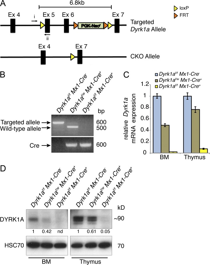
Conditional inactivation of the Dyrk1a gene. (A) Exons 5 and 6 were floxed in the targeted allele and excised in the conditional knockout (CKO) allele. (B) PCR from thymocyte genomic DNA was performed 2 wk after pI:pC treatment using the indicated primers in A (i and ii) and assessing the presence or loss of the targeted allele in Dyrk1af/f Mx1-Cre−, Dyrk1af/w Mx1-Cre+, and Dyrk1af/f Mx1-Cre1+,mice. (C) Dyrk1a mRNA expression measured by qRT-PCR using primers within the excised gene segment in bone marrow and thymus for the indicated mice. (D) Western blot shows DYRK1A protein expression in bone marrow and thymus after loss of 0, 1, or 2 Dyrk1a alleles. Densitometry values were normalized to HSC70. Loss of DYRK1A expression was verified for all subsequent experiments by qPCR, Western blot, or both. Data are derived from 1 litter and are representative of over 20 cohorts that were analyzed by RT-PCR and/or Western blot.

