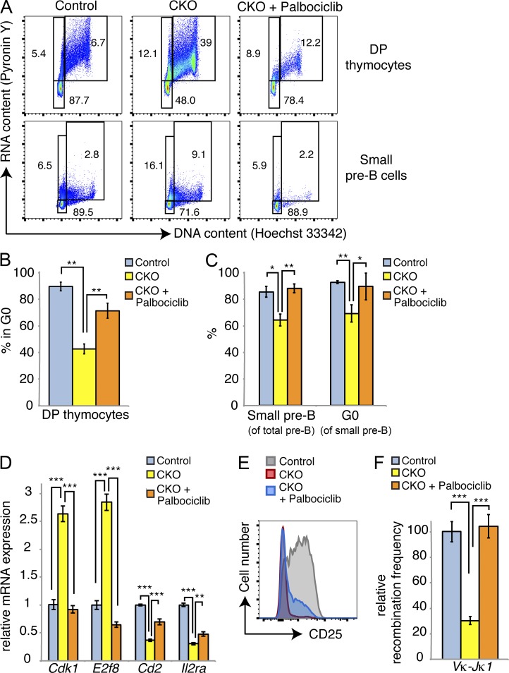
Figure 10.
Inhibition of CDK4/6 can rescue cell cycle exit and differentiation in Dyrk1a-defcient cells. (A) Mice of the indicated genotypes were treated with vehicle or 150 mg/kg Palbociclib (oral gavage) for 1 wk after completion of pI:pC treatment. Cell cycle analysis of DP thymocytes and bone marrow small pre–B cells was assessed. Flow cytometry plots represent two independent experiments, each with two to three mice per condition. (B and C) Quantifications of mean quiescent DP thymocyte and small pre–B cell populations are shown; n = 2–3 mice per genotype; error bars depict SD. Data represent two independent experiments, each with two to three mice per condition. (D) qRT-PCR analysis was performed for the indicated transcripts in FACS-purified small pre–B cells from the mice shown in B and C. Data show two pooled mice per condition; error bars depict SD of triplicate wells. Data are representative of two independent Palbociclib treatment experiments. (E) Flow cytometry plot depicts surface CD25 expression on bone marrow small pre–B cells from a representative mouse from each condition as indicated. Data represent two independent experiments, each with two to three mice per condition. (F) Light chain recombination frequencies were measured by qPCR from genomic DNA in FACS-purified bone marrow small pre–B cells from the indicated mice; each sample was pooled from 2 mice; data represent two independent experiments. *, P < 0.05; **, P < 0.01; ***, P < 0.001.

