Figure 1.
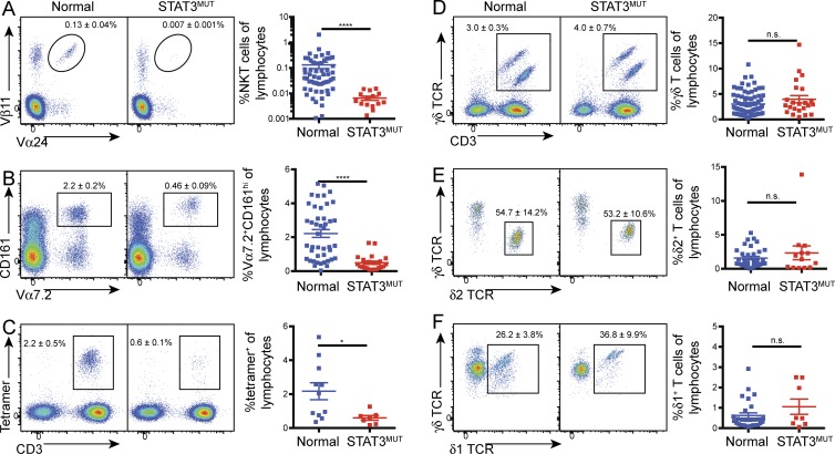
Mutations in STAT3 result in decreased NKT and MAIT cell numbers. (A–F) PBMCs from normal controls or STAT3 mutant patients (STAT3MUT) were stained for iNKT cells (TCRVα24+ Vβ11+; A), MAIT cells [CD3+Vα7.2+ CD161+ (B); CD3+ cells binding MR1–rRL-6-CH2OH tetramers (C)], and total γδ T cells (D), as well as δ2+ (E) and δ1+ (F) subsets. Representative staining of total lymphocytes (A, C, and D), CD3+ cells (B), or γδ T cells (E and F) is shown on the left. Numbers represent mean percentage (±SEM) of lymphocytes (A–D) or γδ T cells (E and F). Graphs show combined data with each symbol representing a single control (n = 11–78) or patient (n = 7–23); error bars indicate SEM; *, P < 0.05; ****, P < 0.0001.

