Figure 2.
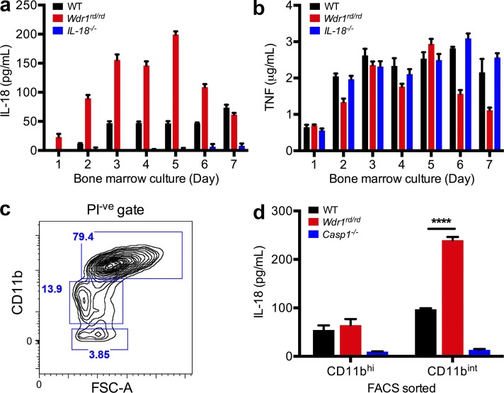
LPS-induced IL-18 secretion from Wdr1rd/rd monocytes, not macrophages. (a and b) BM from WT, Wdr1rd/rd, or IL-18−/− mice was cultured in L929 cell-conditioned medium for 1–7 d. 105 cells from day 1–7 cultures were incubated with 1 µg/ml LPS. 48 h after stimulation, culture supernatants were harvested and assayed by ELISA for IL-18 (a) and TNF (b). Error bars represent SEM of two technical replicates of two biological duplicates. (c and d) BM from WT, Wdr1rd/rd, or caspase-1−/− mice was cultured in L929 cell-conditioned medium for 4 d. (c) Propidium iodide negative (PI-ve) and CD11bhi or CD11bint/low populations indicated by blue boxes were sorted by flow cytometry. FSC-A, forward scatter A. (d) 5 × 10⁴ cells were treated with 1 µg/ml LPS for 48 h. Secreted IL-18 was measured as in panel a. Error bars represent SEM of three technical replicates of two biological duplicates. ****, P < 0.0001 by unpaired t test. All data are representative of two independent experiments.

