Figure 7.
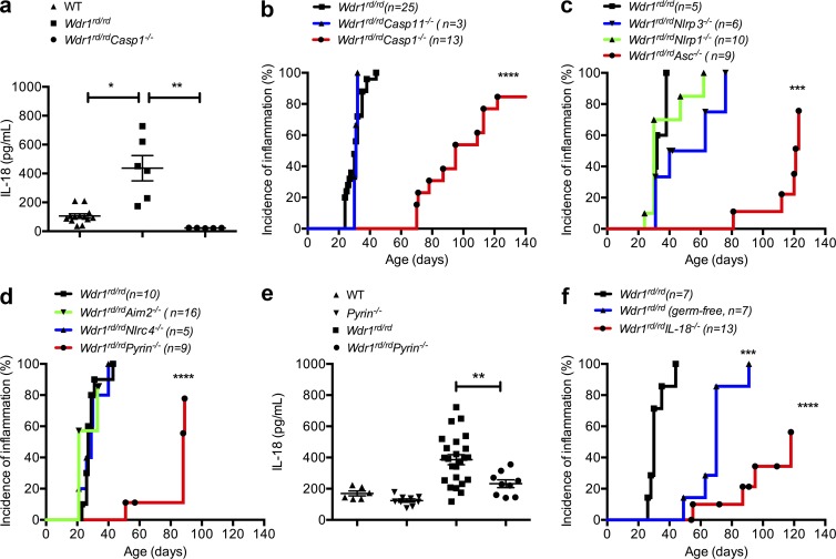
Pyrin-dependent IL-18 secretion and inflammation in Wdr1 mutant mice. (a) Serum IL-18 from WT, Wdr1rd/rd, and Wdr1rd/rdCasp1−/− mice. n = 5–13. *, P < 0.05; **, P < 0.01 by unpaired t test. (b) Incidence of inflammation from Wdr1rd/rd, Wdr1rd/rdCasp1−/−, and Wdr1rd/rdCasp11−/− mice. ****, P < 0.0001 by Gehan-Breslow-Wilcoxon test. (c) Incidence of inflammation from Wdr1rd/rd, Wdr1rd/rdNlrp1−/−, Wdr1rd/rdNlrp3−/−, and Wdr1rd/rdAsc−/− mice. ***, P < 0.001 by Gehan-Breslow-Wilcoxon test. (d) Incidence of inflammation from Wdr1rd/rd, Wdr1rd/rdNlrc4−/−, Wdr1rd/rdAim2−/−, and Wdr1rd/rdPyrin−/− mice. ****, P < 0.0001 by Gehan-Breslow-Wilcoxon test. (e) Serum IL-18 from WT, Pyrin−/−, Wdr1rd/rd, and Wdr1rd/rdPyrin−/− mice. (f) Wdr1rd/rd mice were rederived in a gnotobiotic facility to establish any potential role for commensal microbes in the inflammatory pathology caused by a loss of Wdr. This is compared with conventionally housed Wdr1rd/rd and Wdr1rd/rdIL-18−/− mice. ***, P < 0.001; ****, P < 0.0001 by Gehan-Breslow-Wilcoxon test. Except for disease incidence, data are representative of two to four independent experiments. n = 5–25. Error bars represent means ± SEM.

