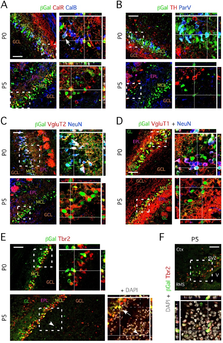
Fig 2. ND1-expressing cells located in the mitral cell layer transiently express markers associated with glutamatergic neuron phenotype.
A- Immunohistochemistry of sagittal OB section of ND1+/LacZ mice aged P0 shows β-Gal+ cells co-expressing CalR and CalB; white arrowhead pinpoint at a β-Gal+/CalR+/CalB+ triple labeled cell. β-Gal+/CalR+ cells are found in the mitral cell layer at P5; co-expression with CalB is lost at P5. Right panels represent higher magnification images of framed areas with orthogonal projections. B- Immunohistochemistry of sagittal OB section of ND1+/LacZ mice aged P0 shows β-Gal+ cells co-expressing ParV. Co-expression with ParV is lost at P5. TH is never found co-expressed by β-Gal+ cells and only locates in β-Gal- cells that migrate towards the forming glomeruli. Right panels represent higher magnification images of framed areas. C- Immunohistochemistry of sagittal OB section of ND1+/LacZ mice aged P0 shows β-Gal+ cells co-expressing VGluT2 and NeuN. NeuN expression is lost at P5 while the expression of VGluT2 persists in β-Gal+ cells. Right panels represent higher magnification images of framed areas. D- Immunohistochemistry of sagittal OB section of ND1+/LacZ mice aged P0 shows β-Gal+ cells co-expressing VGluT1 and NeuN. NeuN expression is lost at P5 while, as VGluT2, VGluT1 expression persists in β-Gal+ cells. Right panels represent higher magnification images of framed areas. E- Immunohistochemistry of sagittal OB section of ND1+/LacZ mice aged P0 shows β-Gal+ cells co-expressing Tbr2. At P5, Tbr2-/β-Gal+, Tbr2+/β-Gal- and Tbr2+/β-Gal+ (pinpointed by a white arrowheads) are observed in the granule layer and forming glomeruli. Right panels represent higher magnification images of framed areas and include DAPI staining (in grey). The white arrow pinpoints at a Tbr2+/β-Gal+ cell in the MCL. F- Tbr2-/β-Gal+, Tbr2+/β-Gal- and Tbr2+/β-Gal+ locate in the SVZ of P5 animals. Lower panel represent a higher magnification image of the framed area and includes DAPI staining (in grey). A- to F-: SVZ = subventricular zone, MCL = mitral cell layer, RMS = rostral migratory stream, GCL = granule cell layer, GL = glomeruli, EPL = external plexiform layer, V = ventricle, Ctx = cortex. A- to E-: Images represent areas from middle sagittal sections of the anterior OB. Scale bars: 50 μm (A-F).

