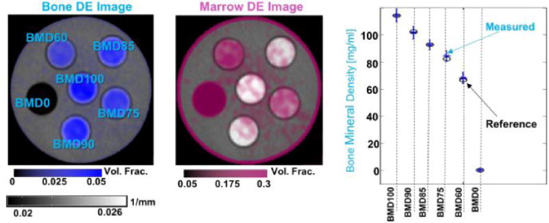
Figure 2.

Left: DE map of bone volume fraction superimposed on a single energy reconstruction of the DE phantom. Center: DE map of marrow volume fraction. Left: Measured (blue markers) and reference (black circles) BMD values for the six inserts of the DE phantom.
