
Figure 2. RNAi-mediated knockdown of dFz2 function in Protocerebral Anterior Medial (PAM) dopaminergic neurons causes flight deficits.
(A) Expression pattern of THGAL4 in the anterior and posterior regions of the brain are shown. Dotted line markings show the neuronal clusters. PAM: protocerebral anterior medial; PAL: protocerebral anterior lateral; PPM1, PPM2, PPM3: protocerebral posterior medial 1, 2, and 3; PPL1, PPL2: protocerebral posterior lateral 1 and 2. (B) Percentage flight times of heterozygous GAL4 controls (gray bars) and GAL4-specific knockdown of dFz2 (red bars). Knockdown of dFz2 in PAM-expressing GAL4 individuals (THC1GAL4, NP6510GAL4, R58E02GAL4) resulted in significantly reduced flight times when compared to their respective GAL4 controls (*p < 0.001, Mann–Whitney U-test). (C) Durations of rhythmic action potentials recorded from the DLMs of air-puff stimulated tethered flies. Average Spike durations were reduced upon expression of dFz2 RNAi in NP6510GAL4 and R58E02GAL4 as compared to GAL4s controls (*p < 0.001, Mann–Whitney U-test). (D) Expression of THC1GAL4, THF2GAL4, NP6510GAL4, and R58E02GAL4 in the PAM neuronal cluster is shown. Except THF2GAL4, all other GAL4s express in dopaminergic PAM neurons. Expression was analyzed from 10 brain hemispheres.

Figure 2—figure supplement 1. Electrophysiological traces showed reduced firing upon knockdown of dFz2.
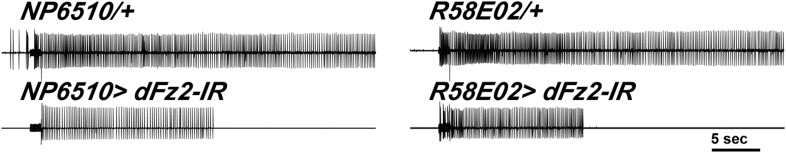
Figure 2—figure supplement 2. Expression of dFz2 in PAM dopaminergic neurons.

Figure 2—figure supplement 3. Expression of TH in PAM dopaminergic neurons during development.
