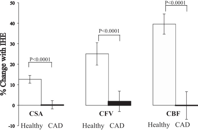
Fig. 3.

Coronary responses to IHE in healthy subjects and patients with CAD. Summary coronary artery vasoreactive changes to IHE stress (as %baseline values) for CSA, coronary blood velocity, and coronary blood flow (CBF) detected by MRI in healthy subjects (white bars) and patients with CAD (black bars).
