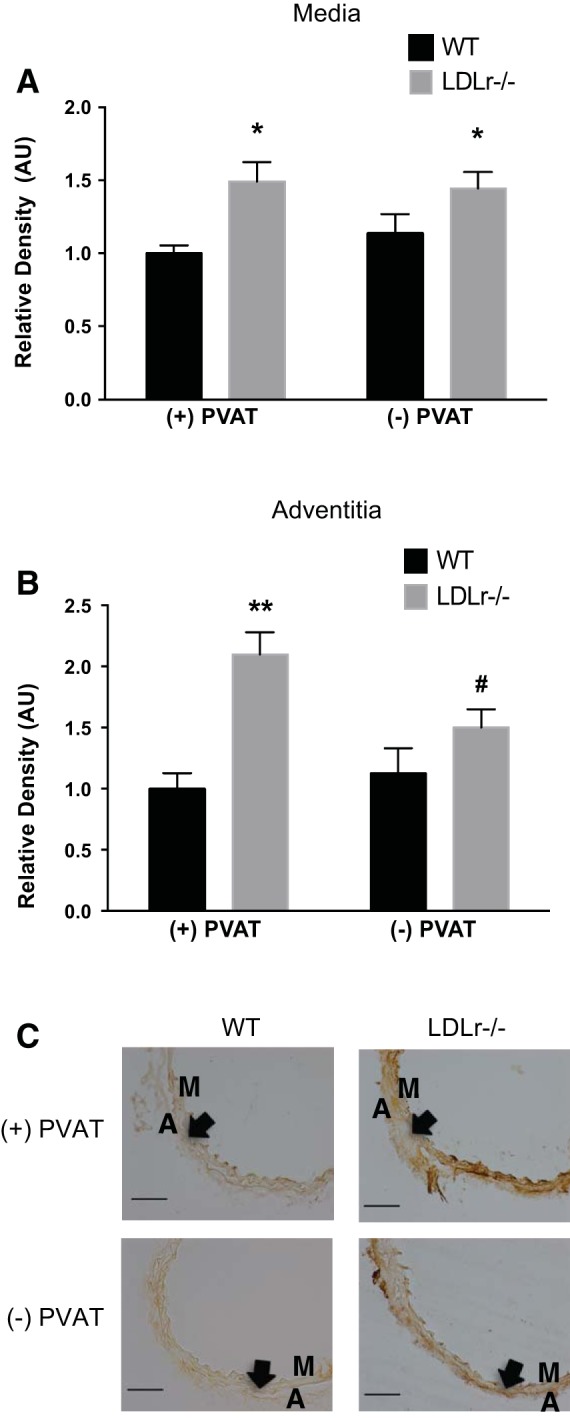
Fig. 5.

Effects of aortic PVAT on aortic AGEs. A and B: medial (A) and adventitial (B) AGEs in histological cross-sections (C) of aortic segments cultured for 72 h in the presence or absence of PVAT from WT and LDLr−/− mice. Values are means ± SE; n = 6–8 mice/group. *P < 0.05 for main effect of effect of strain in the medial layer; **P < 0.05 vs. WT + PVAT, WT − PVAT, and LDLr−/− − PVAT; #P < 0.05 vs. WT mice with PVAT. Arrows denote the medial-adventitial border. Scale bar = 100 μm.
