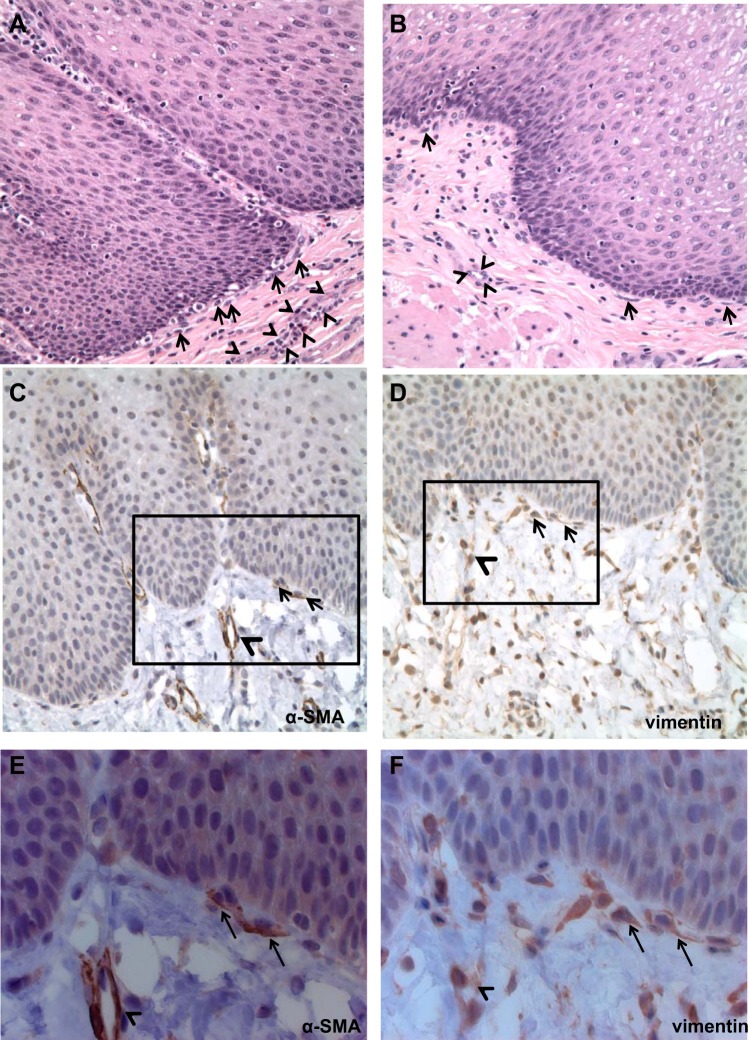
Fig. 1.
Immunostaining of normal human esophageal stroma. Hematoxylin and eosin staining was performed on full-thickness sections of normal esophagus. Examination of the stroma (n = 4; A and B) demonstrates a heterogeneous population with spindle-shaped cells near the basement membrane of the squamous epithelium (arrows). Blood vessels are delineated by arrowheads. Magnification, 400×. Immunostaining demonstrates α-smooth muscle actin (α-SMA; n = 2; C and E) expression in endothelial cells forming blood vessels (arrowheads) and in spindle-shaped cells adjacent to the squamous epithelium (arrows). Immunostaining for vimentin (n = 2; D and F) demonstrates expression in several stromal cells, including endothelial cells forming blood vessels (arrowheads) and in spindle-shaped cells near the squamous epithelium (arrows; A and B, 200×; C and D, 400×). The black boxes in C and D depict areas shown in E and F (630×).

