Fig. 13.
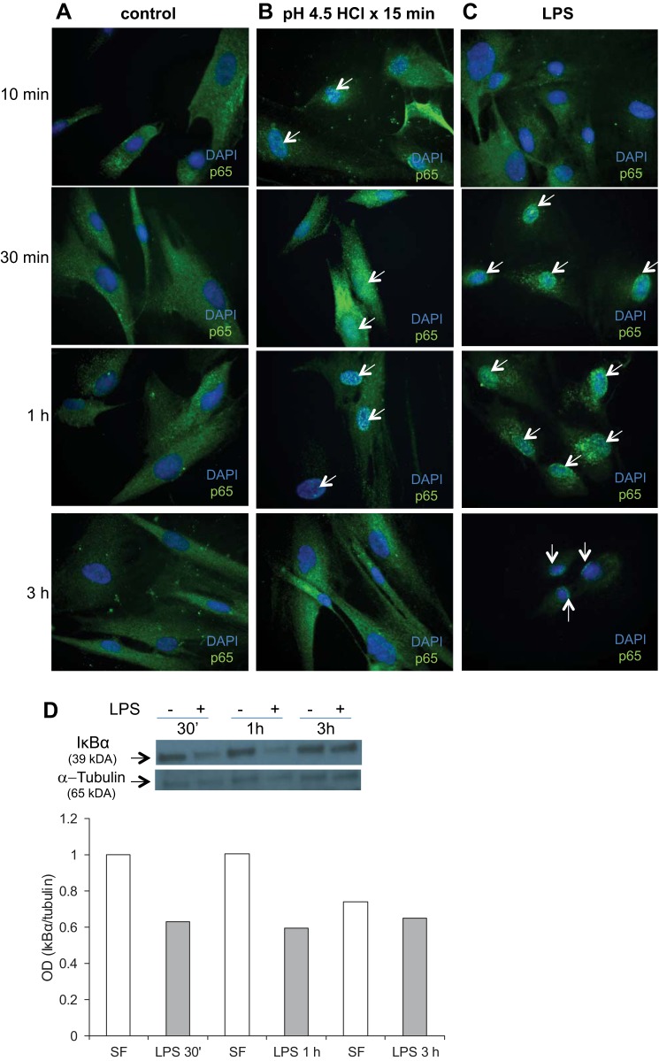
Activation of the NF-κB pathway in acid- and LPS-treated esophageal myofibroblasts. Primary cultures of esophageal myofibroblasts were grown in chamber slides in serum-free myofibroblast media and fixed at 10 min, 30 min, 1 h, and 3 h. These cells served as controls (A). Esophageal myofibroblasts treated with pH 4.5-acidified media for 15 min were then recovered in serum-free myofibroblast media for 10 min, 30 min, 1 h, and 3 h and fixed (B). Cells cultured in LPS were fixed at similar time points (C). Cells were immunostained for p65 with DAPI counterstain. Untreated cells cultured in serum-free myofibroblast media demonstrate cytoplasmic p65 expression at all times (A, green cytoplasm, blue DAPI-stained nucleus). Cells treated with acidified media (B) demonstrate green specks in the nucleus, consistent with nuclear translocation beginning at 10 min (arrows), with more intense nuclear staining of p65 observed at 30 min and 1 h (arrows) compared with untreated cells. At 3 h, esophageal myofibroblasts treated with acidified media demonstrate predominantly cytoplasmic p65 staining. Magnification, 400×. Cells treated with LPS (C) demonstrate green nuclear specks with minimal cytoplasmic staining, consistent with near-complete p65 nuclear translocation, beginning at 30 min and persisting at 3 h. D: cell lysates were harvested from primary cultures grown in serum-free myofibroblast media (SF) and from cultures treated with LPS and harvested at indicated time points after treatment (30 min, 1 h, and 3 h). Immunoblots for inhibitor of NF-κBα (IκBα) were performed on protein harvested from untreated cells and cells treated with LPS. Blots were stripped and reprobed for tubulin as loading control. At each time point, a decrease in IκBα expression is observed after treatment with LPS (top). Relative quantification of IκBα bands was achieved by normalizing the densitometry of the band to the tubulin-loading control using ImageJ software.

