Fig. 4.
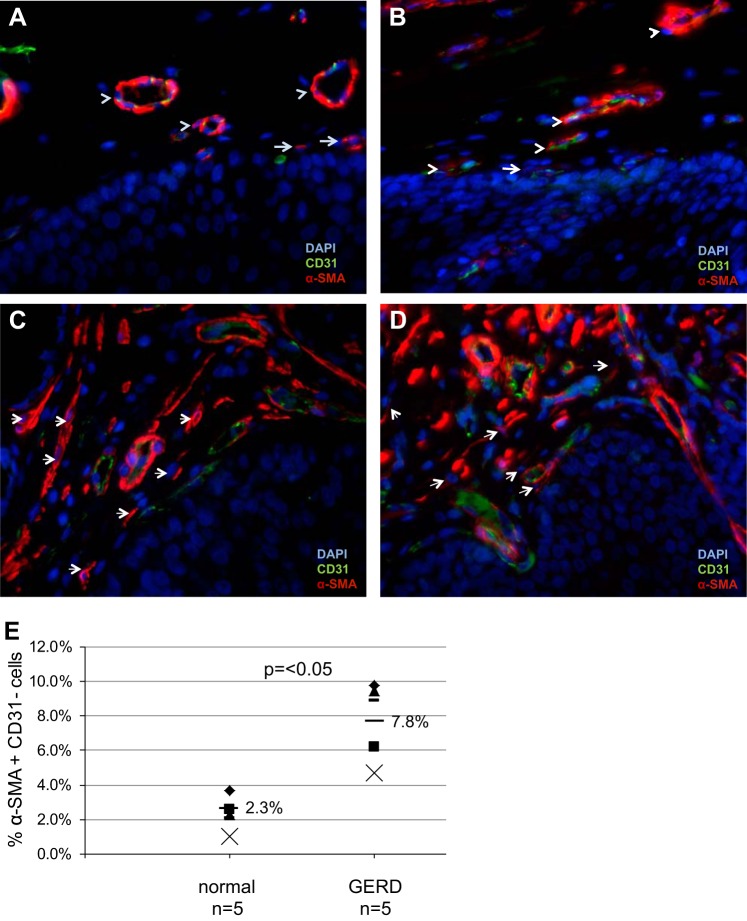
Expression of α-SMA-positive, CD31-negative stromal cells in normal and GERD esophagus. α-SMA (TRITC, red) and CD31 (FITC, green) immunostaining was performed on normal esophagus (A and B) and GERD esophageal (C and D) biopsies to distinguish myofibroblasts (α-SMA+CD31−) from endothelial cells (α-SMA+CD31+). Images were viewed with an immunofluorescent microscope. In normal esophagus (A and B), coexpression of α-SMA and CD31 is readily observed in endothelial cells forming blood vessels (arrowheads). Occasional α-SMA+CD31− myofibroblasts (arrows) are also observed. In GERD biopsies (C and D), α-SMA+CD31− myofibroblasts are observed more frequently (arrows). Representative images of 2 GERD biopsies are shown; magnification, 400× oil. E: quantification of SMA+CD31− cells was performed. GERD esophagi (n = 5) had an increase in the percentage of subepithelial SMA+CD31− myofibroblasts compared with normal esophagus (mean 7.8 vs. mean 2.3%, P < 0.05).

