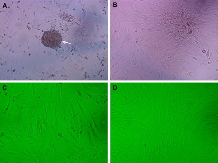
Fig. 6.
Primary cultures of human esophageal stromal cells. Within hours of plating, a suspension of partially floating cells, loosely adherent to the plate bottom, is observed (A; arrow; magnification, 100×). Within 24–48 h, the mixed cell suspension flattens out and adheres to the plate bottom, followed by an outgrowth of spindle-shaped cells (B). Representative images of human stromal cell cultures at low (C) and high (D) confluence are shown at 100× magnification.

