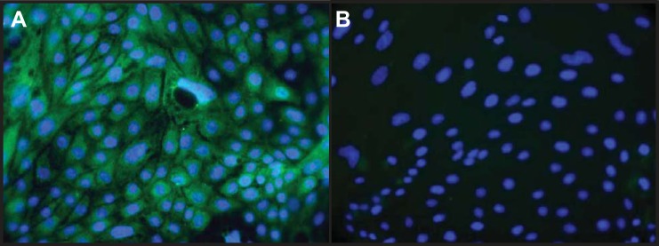
Fig. 1.

Immunocytochemical identification of cultured renal proximal tubule cells (RPTCs). A: fluorescence image of RPTCs derived from kidneys from control animals stained with an antibody to anti-Na+-glucose cotransporter (SGLT)2, a marker for RPTCs (fluorescein, green), and the nuclear marker 4′,6-diamidino-2-phenylindole (DAPI; blue). B: control incubations where the primary antibody was omitted. Magnification: ×400.
