Fig. 7.
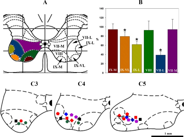
Changes in GAD65/67-immunopositive cells after anti-GAD65/67 siRNA injection into the C4 ventral horn, and location of respiratory interneurons. A: C4 transverse spinal section taken from Paxinos and Watson stereotaxic rat brain atlas (55), where from VII to IX are Rexed's layers in C4 ventral horn and M, L, and VL are medial, lateral, and ventrolateral layer subdivisions, respectively. Grid spacing is 1 mm. B: normalized (% of saline-microinjected control set of animals) number of GAD-65/67-positive cells after anti-GAD65/67 siRNA injection into the C4 ventral horn (colors of bars corresponded to layers in A); asterisks (*) show significant (P < 0.05) difference in GAD-65/67 expression compared with control. C3–C5: distribution of respiratory interneurons recorded from C3–C5 ventral horn: squares, inspiratory with extended expiratory activity (I+E; n = 6); circles, decrementing inspiratory with extended expiratory activity (I-Dec+E; n = 5); pentagons, augmenting expiratory (E-Aug; n = 7); diamonds, slow decrementing expiratory (E-Dec; n = 5). GAD65/67-positive (n = 8) cells are indicated in red and GAD65/67-negative cells are indicated by other solid colors (I+E, black squares; I-Dec+E, green circles; E-Aug, violet pentagons; E-Dec, blue diamonds), respectively. Scale bar is 1 mm.

