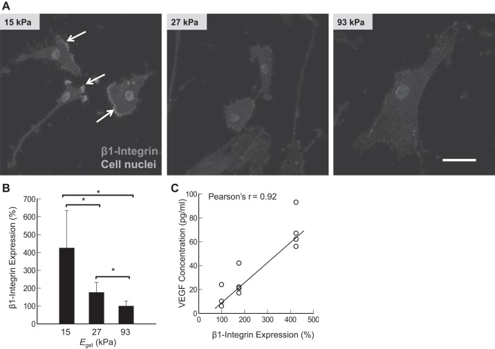
Fig. 6.
Effects of elastic moduli of gels (Egel) on integrin-β1 expression. A: confocal images of cells stained for the integrin-β1 (red color). White arrows indicate the leading edge of ASMCs. B: inverse dependency of integrin-β1 on Egel. C: correlation between the integrin-β1 expression level and the VEGF secretion level. Bars and error bars represent the means ± SD. Scale bar = 50 μm. In these experiments, human ASMCs were seeded on the collagen-conjugated polyacrylamide gels at a density 1,000 cells/cm2. Integrin-β1 expression was detected by staining with anti-β1-integrin antibodies at 24 h postseeding. *P < 0.05, statistically significant difference of values between conditions in B.

