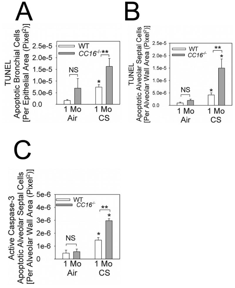
Figure 6. Apoptosis rates are increased in bronchial epithelial and alveolar septal cells in CC16-/- mice exposed to CS.

Terminal deoxynucleotidyl transferase dUTP nick end labeling (TUNEL) staining and immunostaining for active (cleaved) caspase-3 were performed on formalin-fixed lung sections from WT vs. CC16-/- mice exposed to air or CS for 1 month. In A, TUNEL-positive cells were quantified in large and medium-sized airways from 3 air-exposed WT or CC16-/- mice and 4 CS-exposed WT or CC16-/- mice. In B, TUNEL-positive alveolar septal cells were counted and counts were normalized to unit area of alveolar wall in 4 air-exposed WT or CC16-/- mice and 4 CS-exposed WT or CC16-/- mice. In C, alveolar septal cells that stained positively for active (cleaved) caspase-3 were counted and counts were normalized per unit area of alveolar wall in 4 mice per experimental condition. In A-C, data are means ± SEM; asterisk indicates P < 0.05 compared with air-exposed mice belonging to the same genotype; ** P < 0.05.
