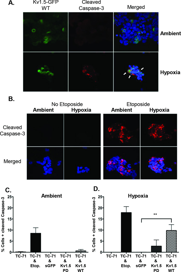
Figure 6. The Kv1.5-WT channel mediates cell death through caspase-3 activation.
(A) Immunocytochemistry and confocal microscopy of Kv1.5-WT expressing TC-71 cells shows induction of caspase-3 cleavage in Kv1.5+ (GFP+) cells (Indicated by white arrows) following exposure to hypoxia. No significant cleavage is detected in Kv1.5+ cells in ambient conditions. (B) Cleaved caspase-3 staining is not detected in parent TC-71 cells in either ambient or hypoxic conditions. To serve as a positive control for cleaved caspase-3 staining, cells were exposed to 4 µg Etoposide. This exposure resulted in robust induction of caspase-3 cleavage in both ambient and hypoxic conditions. Quantification of cells with cleaved caspase-3 is shown for cells in A & B, under ambient (C) (21% O2) and hypoxic (1%O2) (D) conditions. ** p<0.005 (mean ± SEM, n=2–3. 150–400 cell nuclei were counted for each condition).

