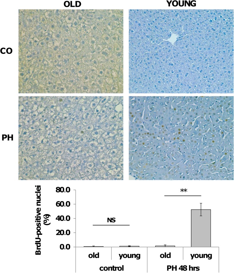
Fig. 2.
Hepatocyte proliferation after PH in old and young livers. a Representative photomicrographs showing BrdU incorporation of hepatocytes in old and young mice after 2/3 PH (sections counterstained with hematoxylin, X200). Mice were subjected to PH and sacrificed 48 h later. All mice were given BrdU (1 mg/ml) in drinking water until the time of sacrifice. CO controls. b Labeling index of hepatocytes from old and young mice subjected to 2/3 PH. At least 2000 hepatocyte nuclei per liver were scored. LI was expressed as number of BrdU-positive hepatocyte nuclei/100 nuclei. Results are expressed as means ± SE of four to five mice per group **Significantly different for p < 0.001; NS not significant

