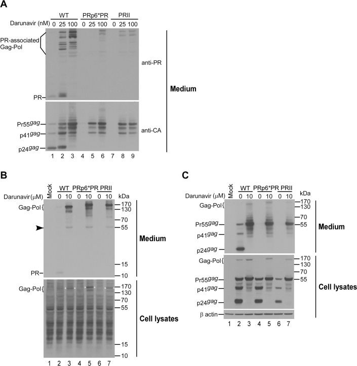
Fig 2. Incorporation of PR and PR-associated Gag-Pol into virus particles.
293T cells were transfected with 10 μg of designated constructs. At 4 h post-transfection, equal amounts of cells were placed on three (panel A) or two (panel B) dish plates, and either left untreated or treated with darunavier at the indicated concentrations. At 48 h post-transfection, cells and supernatants were collected and subjected to Western immunoblotting. PR and PR-associated Gag-Pol were probed with an anti-PR serum (upper panels A and B). Faint bands corresponding to Pr55gag likely indicate partial cross-reaction with anti-PR serum (panel B, arrowhead). Membranes were stripped and reprobed with an anti-p24CA monoclonal antibody. Panels B and C are derived from the same blot. Positions of molecular size markers and HIV-1 Gag proteins Pr55, p41 and p24, and PR-associated Gag-Pol proteins are indicated.

