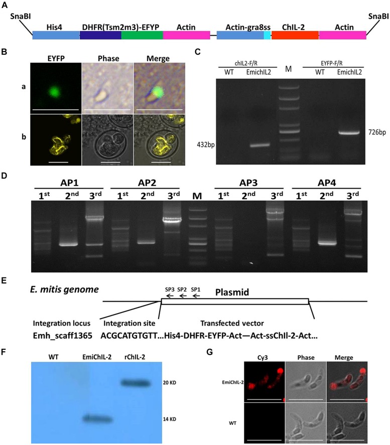
FIGURE 1.
Construction of transgenic Eimeria mitis expressing secreted chicken IL-2 (ChIL-2). (A) Schematic of double-cassette expression vectors. The selection gene [DHFR-Ts-enhanced yellow fluorescent gene (EYFP)] and ChIL-2 were driven by the histone 4 and actin promoter, respectively. Signal sequences (ss) from T. gondii GRA 8 regulated the secretion of ChIL-2. (B) Both the transiently transfected E. mitis sporozoites (a) and the stable transfected EmiChIL-2 (b) were expressing EYFP. (C) Genomic DNA from EmiChIL-2 was amplified with the primers ChIL-2-F and ChIL-2-R (giving a 432 bp product) to verify the recombination of ChIL-2, and the primers EYFP-F and EYFP-R (giving a 726 bp product) to confirm the recombination of EYFP as a positive control, genomic DNA from wild type E. mitis was used as a control. (D) Genomic DNA from EmiChIL-2 was amplified with arbitrary degenerate primers (AP 1, AP 2, AP 3, and AP 4) and specific primers [SP 1, SP 2, and SP 3 (Table 2)] from histone 4 promoter by thermal asymmetric interlaced PCR, and the products from the third-round PCR were cloned into pEasy-T1 vector for sequencing. (E) One integration site (Emh_scaff1365) was confirmed by BLAST from more than 50 clones in the E. mitis GeneDB. (F) Oocysts antigens extracted from EmiChIL-2 reacted with the poly antibody against ChIL-2 producing a clear band with a size of approximately 14 kd to verify ChIL-2 expression by WB. Recombinant ChIL-2 (with two His 6 tag) and the wild-type E. mitis oocysts antigens were used as a positive and negative control, respectively. (G) EmiChIL-2 sporozoites reacted with the poly antibody against ChIL-2 to confirm the localization of ChIL-2 by IFA, and the wild-type E. mitis sporozoites were utilized as a control. Bar = 10 μm.

