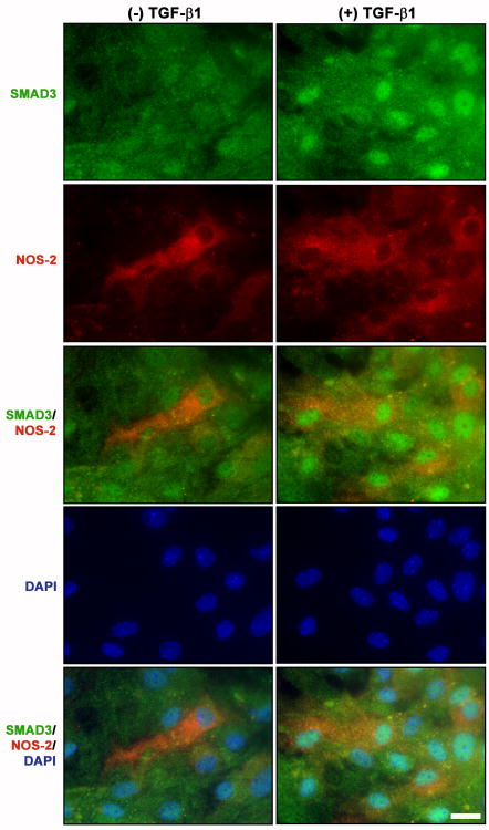
Figure 5. Immunocytochemical assessment of astrocytes exhibiting Smad3 nuclear accumulation and iNOS expression.
Cultures were treated with either vehicle [(-) TGF-β1] or TGF-β1 [(+) TGF-β1; 3ng/ml] for 24 hr prior to the addition of medium containing LPS plus IFNγ (final = 2μg/ml and 3ng/ml, respectively). Eight hr later, cultures were fixed and immunolabeled for SMAD3 (green) and iNOS (red) followed by DAPI counterstaining (blue) to illustrate the number of nuclei per field. Representative photomicrographs (63× magnification) from at least three experiments are shown for each treatment. Scale bar = 20μm.

