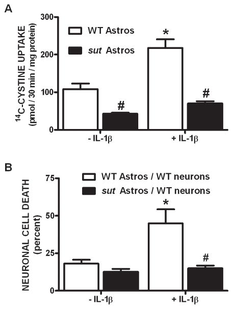
Figure 8. Cystine uptake and hypoxic neuronal cell death are reduced in cultures containing sut astrocytes.
(A) Pure astrocyte cultures (n = 5–6) derived from either wild-type (white bars) or sut mice (black bars) [all cultured w/55 μM β-ME] were treated with vehicle or IL-1β (3 ng/ml) for 20–24 hr after which 14C-L-cystine uptake was determined. Data are expressed as mean ± SEM 14C-L -cystine uptake in pmol/30 min/mg protein. (B) Chimeric mixed cortical cell cultures were obtained by plating wild-type neurons on astrocytes derived from sut mice (black bars). These and control cultures (WT neurons on WT astrocytes; white bars) were treated with 1 ng/ml IL-1β or vehicle for 20–24 hr, washed, and then deprived of oxygen for 5 hr. The percentage of total neuronal cell death was determined 20–24 hr later (n = 4 cultures pooled from two independent experiments). An asterisk (*) indicates a significant within-group difference, while a pound (#) sign indicates a significant between-group difference as determined by a two-way ANOVA followed by Bonferroni’s post hoc test. Significance was set at p < 0.05.

