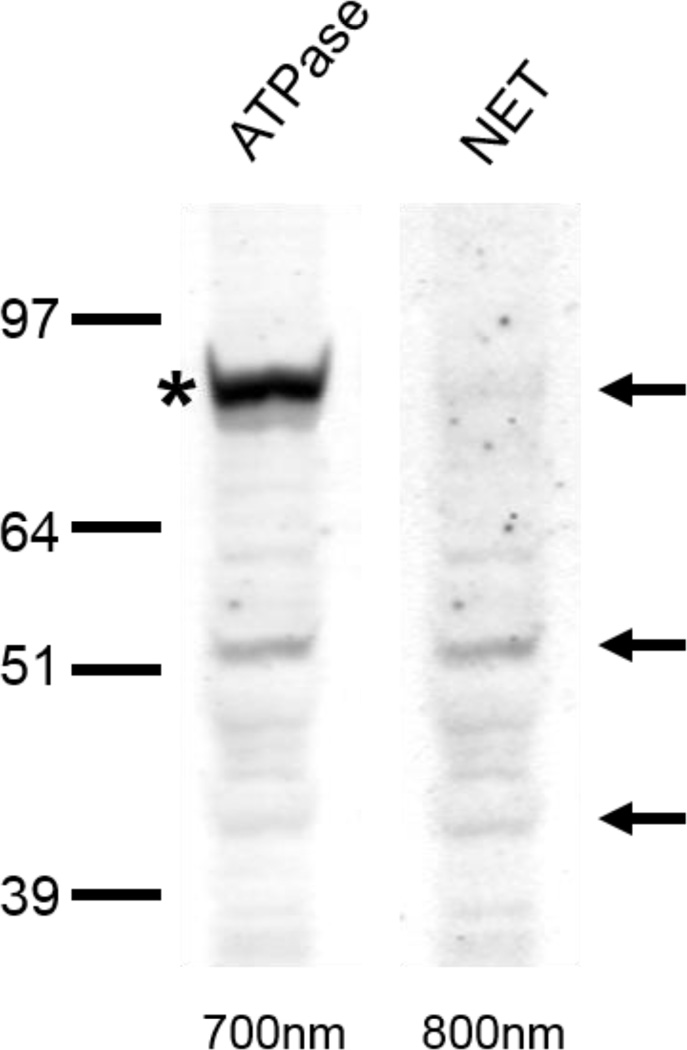
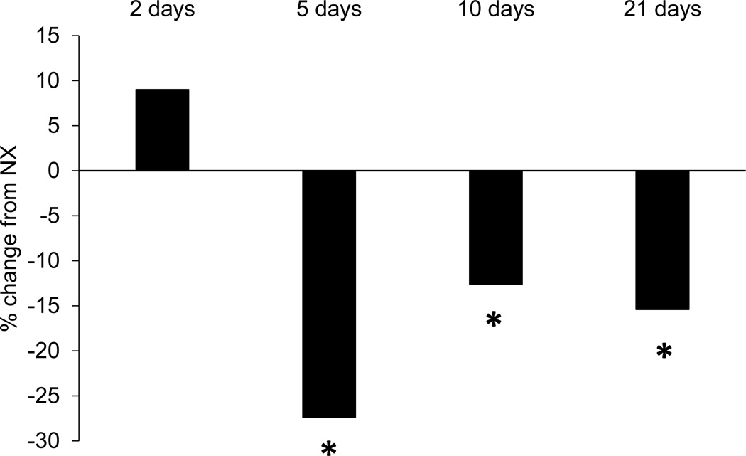
Figure 1.
A) Immunoreactivity of rat pre-frontal cortex to Na, K-ATPase and NET double-labeling. Crude membrane protein fractions were isolated from the pre-frontal cortex and subjected to LDS-PAGE and western blot. Arrows indicate the 80-, 54- and 46-kDa NET proteins. Asterisk indicates Na, K-ATPase. Due to secondary-secondary antibody interactions, NET bands are visible in both 700nm and 800nm Odyssey Imager channels. B) NET levels in the medial prefrontal cortex of rats that exercised as expressed as a percentage of the levels observed in non-exercising rats. Five, 10, or 21 days of exercise significantly reduced NET levels compared to non-exercising controls. *p<0.05.


