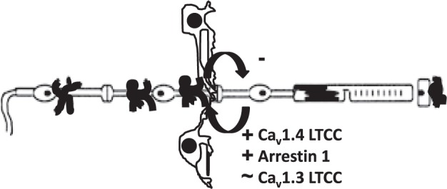
Figure 3.

Simplified schematic representation highlighting the targets studied. Illustration of the retina and support circulations (using conventions in Fig. 1), including the HCs.66–68 In the dark, rod cell membranes are depolarized, resulting in the sustained opening of synaptic LTCCs of the Cav1.4 subtype and presumably Cav1.3 LTCCs in rod nuclei.4–6 Persistent opening of Cav1.4 channels (but not, apparently, Cav1.3 LTCCs) triggers continuous release of the neurotransmitter glutamate, a process regulated by arrestin-1.7,8 When HCs receive glutamatergic input, their membranes depolarize and send (by a yet unclear process) inhibitory signals back to the photoreceptors.9,10 The present data suggest that the target proteins chosen (based on light-based studies ex vivo) also participate in inhibitory signaling in mice in total darkness in vivo when HCs experience a maximum and sustained glutamate exposure.
