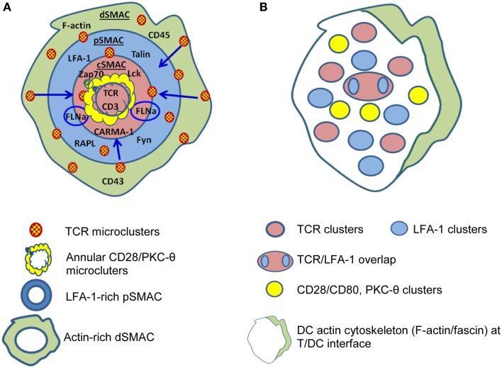
Figure 1.
Differences are apparent between immunological synapses formed by B cells and dendritic cells (DC). (A) B cells, B cell tumors, and lipid bilayers form classical “bulls-eye” immunological synapses. TCR-containing microclusters form in the dSMAC, contain Lck and ZAP70, protein kinase C (PKC-θ), LAT, SLP76 etc., and migrate centripetally through the LFA-1-rich pSMAC to the cSMAC. The cSMAC is segregated into a central CD3hi region where CD3 accumulates and TCR is internalized to resulting in termination of TCR signaling and an outer CD3lo region where the signaling molecules accumulate either in conjugation [annular CD28/PKC-θ conjugates and PKC-θ/filaminA (FLNa) clusters] or separately. (B) DC typically form “multifocal” synapses where TCR-containing clusters are segregated from CD28/PKC-θ containing clusters and no clear “ring” of LFA-1 is formed. TCR signaling stabilizes the multifocal structure, particularly the CD28/PKC-θ containing clusters. A prominent polarization of the DC cytoskeleton is often present at the periphery. Based on Ref. (51, 53, 55–57) (A); (58–60) (B).

