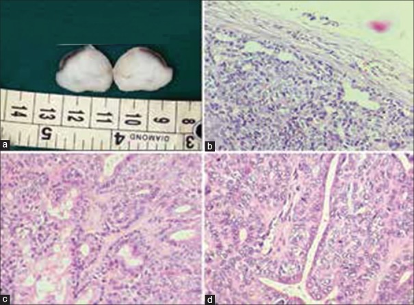
Figure 2.
(a) Gross examination: Tumor with a solid homogenous grey-white lobulated cut surface. (b-d) Histological sections showing basaloid cells with uniform round-oval nuclei, eosinophilic cytoplasm and indistinct cytoplasmic borders arranged in tubulo-trabecular pattern separated by collagenous stroma and peripheral palisading within the nests. (H&E stain, (b) x200, (c) x400, (d) x400)

