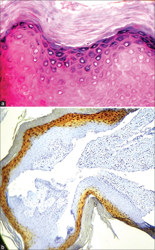
Figure 1.

(a) The histopathological image shows Hyperkeratosis of the human foreskin (H&E stain, x40); (Courtesy: Department of Oral Pathology and Microbiology, Ragas Dental College and Hospital, Chennai). (b) Hyperkeratosis without epithelial dysplasia showing loricrin positivity in the stratum granulosum of Human foreskin (IHC stain, x10); (Courtesy: Department of Oral Pathology and Microbiology, Ragas Dental College and Hospital, Chennai)
